
Labeled Pictures Of The Human Heart koibana.info Heart anatomy
Browse 8,896 human heart anatomy photos and images available, or search for human heart anatomy vector to find more great photos and pictures. Browse Getty Images' premium collection of high-quality, authentic Human Heart Anatomy stock photos, royalty-free images, and pictures.

Image Of The Heart Labeled
ISSN 2534-5079 This interactive atlas of human heart anatomy is based on medical illustrations and cadaver photography. The user can show or hide the anatomical labels which provide a useful tool to create illustrations perfectly adapted for teaching.
BIO202Heart Model
Author: Jana Vasković MD • Reviewer: Alexandra Osika Last reviewed: November 03, 2023 Reading time: 12 minutes Recommended video: Anatomy of the heart [10:27] Overview of the anatomy and functions of the heart. Heart (right lateral view)
The Heart Diagram Labeled and Unlabeled Worksheets Heart Etsy
Picture of Heart. The heart is a muscle that pumps blood throughout the body. The heart is located beneath the rib cage slightly to the left of the center of the chest between the lungs and above the diaphragm. A healthy adult heart is approximately the size of a closed fist. The valves and chambers of the heart work together to pump oxygen.
Heart And Labels Drawing at GetDrawings Free download
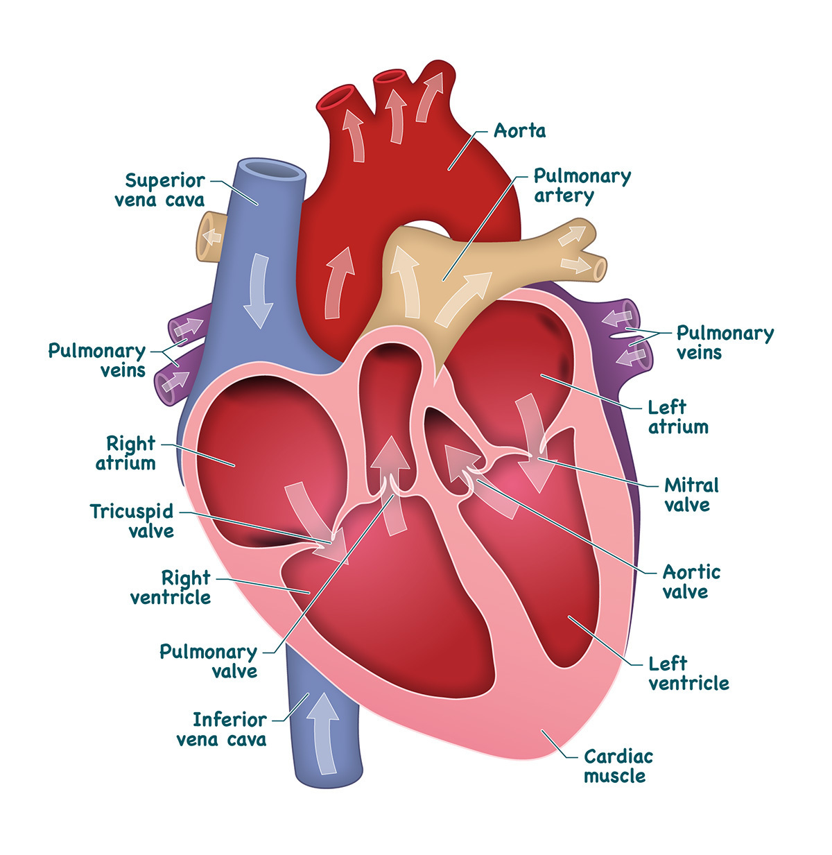
On average, an adult's heart weighs about 10 ounces. Your heart may weigh a little more or a little less, depending on your body size and sex. What are the parts of the heart's anatomy? The parts of your heart are like the parts of a house. Your heart has: Walls. Chambers (rooms). Valves (doors). Blood vessels (plumbing).
Labeled Drawing Of The Heart at GetDrawings Free download
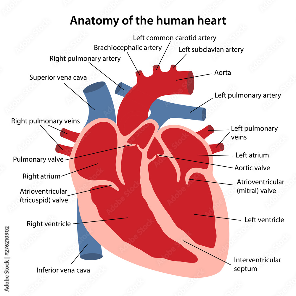
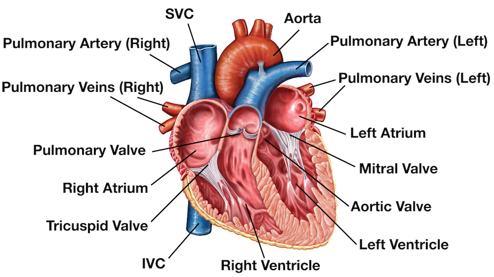
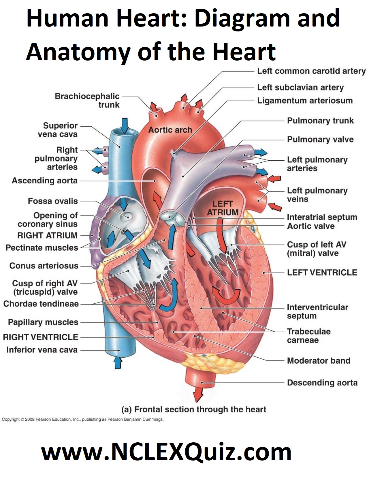
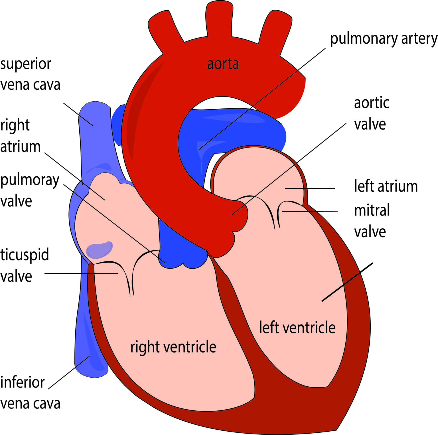
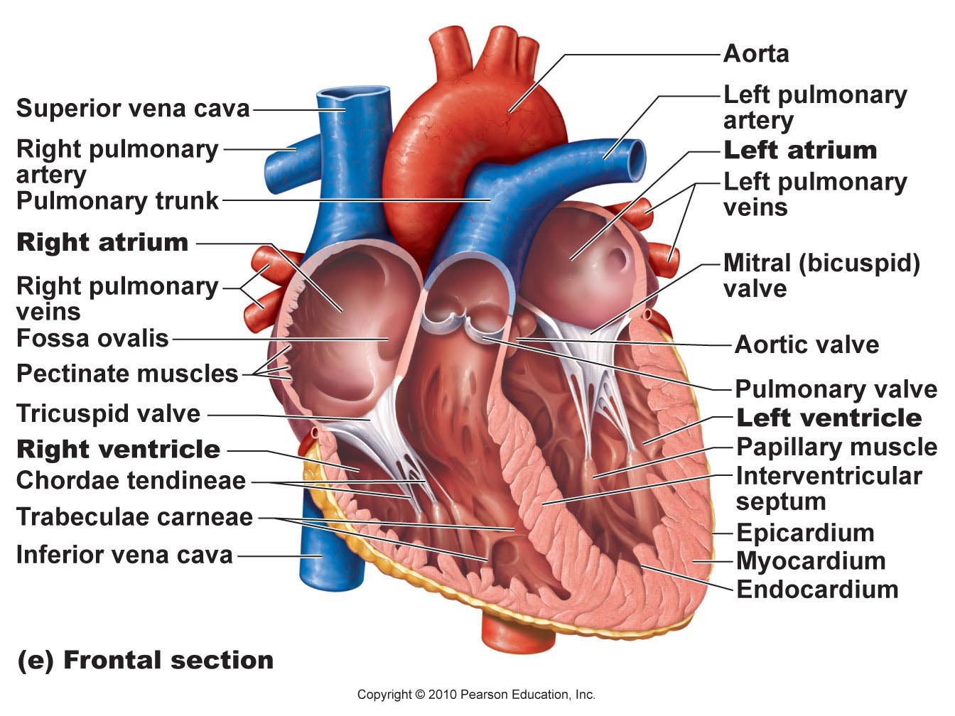
Atria and Ventricles The human heart, comprises four chambers: right atrium, left atrium, right ventricle and left ventricle. The two upper chambers are called the left and the right atria, and the two lower chambers are known as the left and the right ventricles.

Heart Structure and Function Worksheet Teacha!
What is the heart wall made up of? What causes the heart to beat? What are heart sounds? heart, organ that serves as a pump to circulate the blood. It may be a straight tube, as in spiders and annelid worms, or a somewhat more elaborate structure with one or more receiving chambers (atria) and a main pumping chamber (ventricle), as in mollusks.
Heart Diagram Labeled
Heart Pictures, Diagram & Anatomy | Body Maps Human body Circulatory System Heart Heart The heart is a mostly hollow, muscular organ composed of cardiac muscles and connective tissue.
Heart Anatomy Labeled Diagram, Structures, Blood Flow, Function of
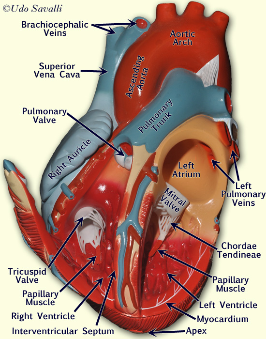
By: Tim Taylor Last Updated: Jul 30, 2020 2D Interactive NEW 3D Rotate and Zoom Anatomy Explorer Heart Aortic Valve Bundle Branches Chordae Tendineae Interventricular Septum Left Atrium Left Auricle Left Ventricle Mitral Valve Papillary Muscles Pulmonary Valve Purkinje Fibers Right Atrium Right Auricle Right Ventricle Tricuspid Valve

How to Draw the Internal Structure of the Heart (with Pictures)
Awesome & High Quality Here On Temu. New Users Enjoy Free Shipping & Free Return. Come and check at a surprisingly low price, you'd never want to miss it.
Human Heart Diagram and Anatomy of the Heart StudyPK
Anatomy The heart is an organ that weighs approximately 350 grams (less than one pound). It's nearly the size of an adult's clenched fist. It's located in the thorax (chest)—between the lungs —and extends downward between the second and fifth intercostal (between the ribs).
On Heart Kardiohirurgija.rs
17,477 human heart diagram stock photos, 3D objects, vectors, and illustrations are available royalty-free. See human heart diagram stock video clips Filters All images Photos Vectors Illustrations 3D Objects Sort by Popular Human heart with blood vessels. 3d illustration Hand drawn illustration of human heart anatomy.

Labeled Pictures Of the Heart Lovely Simple Human Heart Diagram for
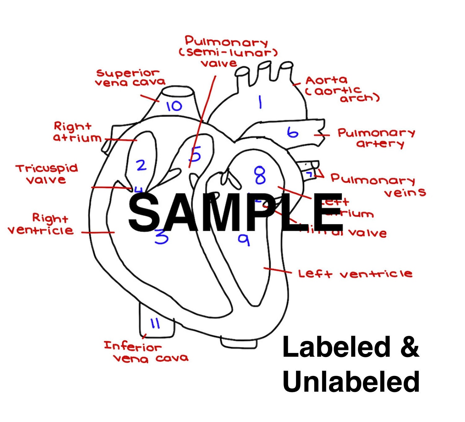
In this interactive, you can label parts of the human heart. and drop the text labels onto the boxes next to the diagram. Selecting or hovering over a box will highlight each area in the diagram. Rights: The University of Waikato Te Whare Wānanga o Waikato
Labeled Drawing Of The Heart at GetDrawings Free download
Location of the Heart. The human heart is located within the thoracic cavity, medially between the lungs in the space known as the mediastinum. Figure 19.2 shows the position of the heart within the thoracic cavity. Within the mediastinum, the heart is separated from the other mediastinal structures by a tough membrane known as the pericardium.
Show me a diagram of the human heart? Here are a bunch! Interactive
Español Your heart is in the center of your chest, near your lungs. It has four hollow chambers surrounded by muscle and other heart tissue. The chambers are separated by heart valves, which make sure that the blood keeps flowing in the right direction. Read more about heart valves and how they help blood flow through the heart.

Heart Anatomy Anatomy and Physiology II
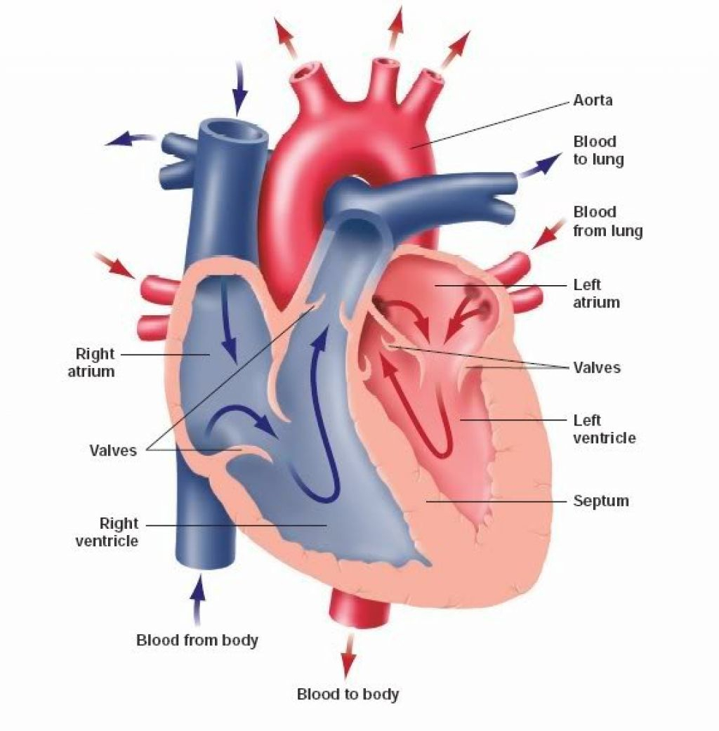
The heart is the organ that helps supply blood and oxygen to all parts of the body. It is divided by a partition (or septum) into two halves. The halves are, in turn, divided into four chambers. The heart is situated within the chest cavity and surrounded by a fluid-filled sac called the pericardium. This amazing muscle produces electrical.